


complete lecture on Cardiovascular System
Cardiovascular System – Complete Explanation
The cardiovascular system, also known as the circulatory system, is one of the most important systems in the human body. It is made up of the heart, blood vessels, and blood. Its main function is to transport oxygen, nutrients, hormones, and other important substances to all the tissues of the body while removing waste products like carbon dioxide and urea. It also helps regulate body temperature, maintain the pH balance of body fluids, and support the immune system by circulating white blood cells.
1. The Heart – Structure and Function
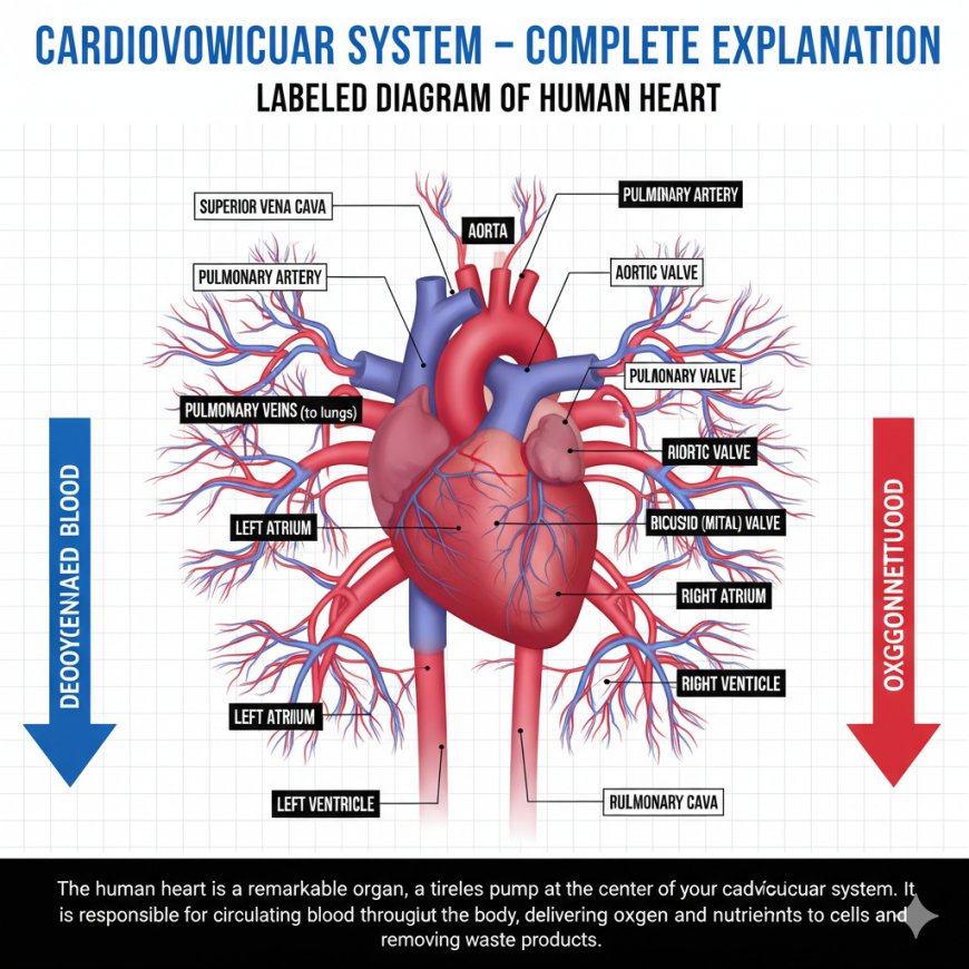
The heart is a strong, hollow, muscular organ about the size of a fist. It is located in the thoracic cavity, between the lungs, and slightly to the left of the midline. The heart is surrounded by a protective covering called the pericardium, which has two layers — the outer fibrous layer and the inner serous layer. The pericardial fluid between these layers reduces friction as the heart beats.
The wall of the heart has three layers:
-
Epicardium – the outer layer that protects and reduces friction.
-
Myocardium – the thick muscular middle layer responsible for pumping action.
-
Endocardium – the smooth inner layer that lines the heart chambers and valves.
The heart has four chambers. The right atrium receives deoxygenated blood from the body through the superior and inferior vena cava. This blood moves into the right ventricle, which pumps it to the lungs through the pulmonary artery for oxygenation. The left atrium receives oxygen-rich blood from the lungs through the pulmonary veins, and this blood then moves into the left ventricle, which pumps it into the aorta to supply the entire body. The left ventricle has thicker walls because it must pump blood under high pressure throughout the body.
To ensure that blood flows in one direction, the heart contains valves:
-
Tricuspid valve (between right atrium and right ventricle)
-
Pulmonary valve (between right ventricle and pulmonary artery)
-
Mitral or bicuspid valve (between left atrium and left ventricle)
-
Aortic valve (between left ventricle and aorta)
These valves open and close according to pressure changes inside the heart during each heartbeat.
2. The Cardiac Cycle
Each complete heartbeat is called a cardiac cycle and has two main phases:
-
Systole – contraction phase (pumping blood out)
-
Diastole – relaxation phase (filling with blood)
During one cycle:
-
Atria contract (atrial systole) to fill the ventricles.
-
Ventricles contract (ventricular systole) to pump blood out of the heart.
-
The heart then relaxes (diastole) and refills for the next beat.
At rest, this cycle lasts about 0.8 seconds, and the heart beats about 70–75 times per minute. The amount of blood pumped by the heart each minute is called cardiac output, calculated as:
[
Cardiac\ Output = Heart\ Rate \times Stroke\ Volume
]
Average cardiac output in an adult is about 5 liters per minute.
3. Electrical Conduction System of the Heart
The heartbeat is controlled by the cardiac conduction system, which produces and conducts electrical impulses to coordinate contraction.
The key components are:
-
Sinoatrial (SA) node – known as the natural pacemaker; located in the right atrium; it starts each heartbeat.
-
Atrioventricular (AV) node – receives impulses from the SA node and delays them slightly to allow the atria to empty before the ventricles contract.
-
Bundle of His – transmits the impulse to the ventricles.
-
Right and left bundle branches – carry the impulse down the interventricular septum.
-
Purkinje fibers – spread the impulse through the ventricular walls, causing them to contract.
This electrical activity can be recorded as an ECG (electrocardiogram):
-
P wave = atrial depolarization (atrial contraction)
-
QRS complex = ventricular depolarization (ventricular contraction)
-
T wave = ventricular repolarization (relaxation)
4. Blood Vessels
The blood vessels form an extensive network throughout the body. There are three main types:
-
Arteries carry blood away from the heart. They have thick, elastic, and muscular walls to handle high pressure. The largest artery is the aorta.
-
Veins carry blood toward the heart. They have thinner walls and contain valves to prevent backflow since the pressure is low.
-
Capillaries are the smallest blood vessels. Their walls are one cell thick, allowing the exchange of oxygen, nutrients, and wastes between blood and body cells.
5. Circulatory Pathways
There are two main circulations:
-
Pulmonary circulation – carries deoxygenated blood from the right ventricle to the lungs and brings oxygenated blood back to the left atrium.
-
Systemic circulation – carries oxygenated blood from the left ventricle to all body tissues and returns deoxygenated blood to the right atrium.
Additionally, the coronary circulation supplies the heart muscle itself with oxygen and nutrients through the right and left coronary arteries.
6. Physiology of Blood Flow and Pressure
Blood pressure (BP) is the force that blood exerts against vessel walls. It depends on cardiac output and total peripheral resistance (resistance of blood vessels).
Normal adult BP is about 120/80 mmHg.
The upper number (systolic pressure) shows pressure during heart contraction, while the lower number (diastolic pressure) shows pressure during relaxation.
Blood flow is also controlled by autoregulation — blood vessels can constrict or dilate depending on the needs of tissues. The autonomic nervous system helps regulate this:
-
Sympathetic nerves increase heart rate and blood pressure.
-
Parasympathetic nerves (vagus nerve) slow the heart rate.
7. Capillary Exchange and Lymphatic System
In capillaries, the exchange of substances occurs mainly by diffusion, filtration, and osmosis.
-
Oxygen and nutrients move from the blood to tissues.
-
Carbon dioxide and wastes move from tissues to blood.
Some fluid leaks out of capillaries into tissues. The lymphatic system collects this excess fluid (now called lymph) and returns it to the bloodstream, preventing tissue swelling (edema).
8. Regulation of Cardiac Function
The heart’s activity is influenced by both intrinsic and extrinsic factors:
-
Intrinsic (Frank–Starling law): The more the heart muscle is stretched by incoming blood (venous return), the stronger the next contraction will be.
-
Extrinsic control: Includes autonomic nerves, hormones (like adrenaline), temperature, and electrolytes.
9. Clinical Importance
Several diseases affect the cardiovascular system:
-
Hypertension (high BP): Increases the risk of heart attack and stroke.
-
Atherosclerosis: Buildup of fatty plaques in arteries that reduces blood flow.
-
Myocardial infarction (heart attack): Blockage of coronary arteries leading to death of heart tissue.
-
Heart failure: When the heart cannot pump enough blood to meet the body’s needs.
-
Arrhythmias: Abnormal heart rhythms due to electrical conduction problems.
10. Summary
The cardiovascular system is a closed-loop transport network that delivers oxygen and nutrients to every cell while removing wastes. The heart acts as the pump, blood vessels act as the pathways, and blood acts as the transport medium. Its function is tightly regulated by electrical, mechanical, and hormonal systems to keep the body’s internal environment stable. A healthy cardiovascular system is essential for life, as every organ depends on a constant, well-regulated blood supply.
What's Your Reaction?
 Like
0
Like
0
 Dislike
0
Dislike
0
 Love
0
Love
0
 Funny
0
Funny
0
 Angry
0
Angry
0
 Sad
0
Sad
0
 Wow
0
Wow
0