


DECRIPTIVE EMBRYOLOGY
Descriptive Embryology deals with the study of an individual organism’s development from fertilization to the fetal stage. It describes each stage of embryonic development in a clear, sequential manner for exam understanding.

DECRIPTIVE EMBRYOLOGY
Basis of Embryo Formation
The study of animal development from the fertilized egg to the formation of all major organ systems is called embryology. All complexity arises from a single egg. There were two views about the origin of complexity in the egg:
1. Preformation:
It tells that the gametes contain all of the elements present in the adult. Most biologists of the seventeenth and eighteenth centuries believed in preformation.
2. Epigenesis:
This theory explains that the egg contains the material from which the embryo is gradually built. This theory became more popular in the mid-eighteenth century.
Aristotle described this theory in fourth century B.C. He named it as creative principle. These principles develop the organism.
Experiments of Roux and Driesch
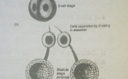
Wilhelm Roux (1888) and Hans Driesch (1892) performed experiments to determine which theory was correct. They allowed a fertilized egg to divide to the two-cell stage.
(a) Roux:
He used amphibian embryos (frogs, toads, salamanders). He killed one of the two cells with a hot needle. The remaining cell formed half embryo.
Roux called this half embryo as hemiembryo. It favors preformation.
(b) Driesch:
He used echinoderm embryos (sea stars, sea urchins, sea cucumbers). He completely separated the divided cells. An entire animal developed from a single cell. It supports epigenesis.
Driesch found that each cell retained the potential to develop entire organism. Biologists now know that Driesch was correct. They believe that the kill
untreated cell.
It was an important turning point in embryology.
FERTILIZATION
Different animals have different mechanisms for bringing opposite sexes together. They also have developed mechanisms to release the eggs and sperm. Similarly, different mechanisms increase the chance of fertilization.

1.
The egg has a gel coat. The gel coat consists of protein, or protein and polysaccharide (mucopolysaccharide). The sperm penetrate into this gel coat.
2.
The sperm of most animals possesses enzymes called sperm lysins. It helps to penetrate this coating. These enzymes are associated with acrosome.
The acrosome is a specialized organelle present at the head of the sperm. Or this enzyme is associated
These enzymes are associated with acrosome.
The acrosome is a specialized organelle present at the head of the sperm. Or this enzyme is associated with the plasma membrane at the anterior tip of sperm.
3.
The sperm touches the gel coat. The acrosome releases lysins. It dissolves the gel coat and forms a pathway for the sperm.
Lysins from many sperm are required to dissolve the gel coat of an egg in humans. Therefore, even 80 million sperm per ejaculation reduces the fertility.
4.
The acrosome of some species releases lysine and reorganizes into an acrosomal process.
Vitelline layer (or zona pellucida) is present just outside the egg plasma membrane. Egg binding protein binding is present on the surface of the
acrosomal process.
It binds the sperm with the attachment molecules of eggs. These attachment molecules are present on the vitelline layer of the plasma membrane of egg.
Acrosomal and egg plasma membranes then fuse.
5.
Other parts of the sperm like mitochondria, centrioles, and flagellum may or may not enter the egg.
EGG ACTIVATION
The fusion of acrosomal and egg membranes is the beginning of egg activation.
Egg activation is a series of biochemical changes in the egg. These changes complete the fertilization and initiates embryonic development. Biologists have studied activation of egg in echinoderms.
Membrane and Cortical Events
1. Some changes occur in the plasma membrane and outer region of the cytoplasm of zygote. This cytoplasm is called the cortex. Thus only a single sperm fertilize the egg. Single-sperm fertilization is necessary. Multiple fertilization cause genetic imbalances. It produces a nonviable embryo.
2.The sperm contact the egg. The microvilli of the plasma membrane of the ovum wrap around a single sperm. The microfilaments in the cytoplasm of egg contract. It draws the sperm into the egg. Ionic changes take place in the plasma membrane within milliseconds. It makes the sperm unresponsive to other sperm. It initiates the formation of a protective envelope around the egg. This protective envelope is called the fertilization membrane.
3. The fertilization membrane forms as granules in the cortex. These granules are discharged into the region between the egg plasma membrane and the vitelline layer. The cortical granules release enzymes. These enzymes loosen the contact of vitelline layer with the plasma membrane.
4. The granules produce space between the :
· Microvilli
· Egg cortex
· Jelly layer
· Fertilization membrane
· Egg plasma membrane
Thus water enters into this space. The vitelline layer lifts off the egg. Proteins of the cortical granules thicken. It strengthens the vitelline layer. All of these reactions are completed in 1 to 2 minutes after fertilization.
5. Other important changes occur in the egg cortex. The cortical layer thickens after sperm penetration. Rotational and sliding movements of the outer egg cytoplasm begin. These cortical changes form a gray crescent on the egg in the amphibians. Gray crescent is present opposite the point of sperm penetration. The gray crescent plays an important role in development.
METABOLIC AND NUCLEAR EVENTS
The nuclear events occur before the nuclear fusion. Post fertilization changes prepare the zygote for the mitotic divisions. Ionic changes raise the intracellular pH. It initiates changes in zygote physiology. DNA replication occurs. The rate of protein synthesis increases. This protein forms the enzymes and structural proteins of the mitotic spindle. Some protein forms structure of chromosomes.
Little mRNA is synthesized in the zygote. Rather, existing maternal mRNA is activated. These mRNA synthesize most of the proteins in early stages of embryonic development. This influence is called maternal dominance. Now egg has two regions.
(a) Animal pole:
The region of the egg containing less yolk, more mitochondria.
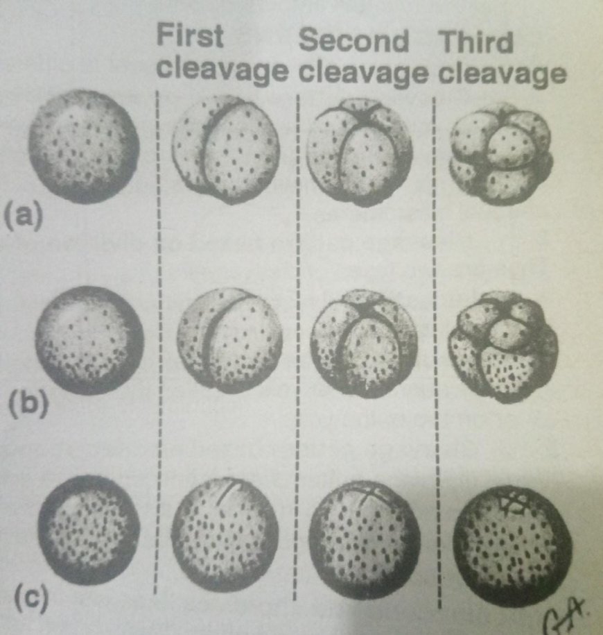
CLEAVAGE
The cell divisions occur during embryonic development are called cleavage.
The cells that cleavage produces are called blastomeres.
The first cell division of the zygote produces two daughter cells. These blastomeres divide at the same time. It produces a four-celled embryo. Later, the divisions become asynchronous and more blastomere are produced.
The embryo does not increase in size during early embryology. Rather, the blastomere become smaller. Thus the proportion of DNA to cytoplasm increases.
QUANTITY AND DISTRIBUTION OF YOLK
The food reserve for the developing embryo containing a mixture of proteins, lipids, and glycogen is called yolk.
Yolk in an egg determines the egg sizes, cleavage patterns, and the length of embryonic periods of animal.
1. Animals with small amounts of yolk (microlecithal):
Its examples are echinoderms (sea stars and their relatives) and amphibians (frogs and their relatives).
They have a brief period of embryological developments. They absorb yolk during this development. After that larva is formed which feed freely.
2. Animal with larger quantities of yolk (megalecithal):
Its examples are reptiles and birds.
They have longer period of embryological development. Thus their embryos have larger quantities of yolk. Animals without yolk or very small yolk (Alecithal):
Its examples are eutherian or placental mammals and sharks. These animals have very long periods of embryological development. The eutherian provide nourishment to embryos through a placenta. The sharks have modification in the female reproductive tract for nourishment of embryo. Thus the eggs of these animals have very small amount of yolk.
CLAEVAGE PATTERNS
Different cleavage patterns are present in different organisms.
1. Cleavage pattern based on amount of yolk:
The yolk influences the cleavage patterns. Some eggs have evenly distributed yolk. Cleavage patterns in these cells produce blastomeres with uniform size. Some eggs have unevenly distributed yolk. Their cleavage patterns produce unequal blastomere
Cleavage pattern based on division of egg:
Thus are two types of cleavages:
* Holoblastic:
Cleavages that completely divide an egg are called holoblastic.
* Meroblastic:
Cleavages that cannot completely divide the embryo due to large quantities of yolk are called meroblastic. These embryos develop around or on top of the yolk.
3. Cleavage pattern based on orientation and fate of blastomeres:
These cleavage patterns are produced due to differences in the orientation of the mitotic spindle during mitosis. It also shows the fate of early blastomeres. The developmental differences show the differences in the evolutionary history of the animal groups.
* Echinoderm and chordates lineages:
In this case, blastomeres are oriented directly over one another. This pattern is present in echinoderms
and chordates.
* Annelids and Arthropods lineage:
In this case, an upper tier of blastomeres is twisted out of line with a lower tier of blastomeres. This pattern is present in the annelids (segmented worms) and arthropods (insects and their relatives).
PRIMARY GERM LAYERS AND THEIR DERIVATIVES
The layers or blocks of embryonic cells which produce tissues and organs are called primary germ layers. The development of these layers from a nondescript form in the early embryo to their form in late embryonic adult stages is called differentiation. There are three layers:
1.Ectoderm:
It gives rise to the outer body wall.
2. Endoderm:
It forms the inner lining of the digestive cavity.
3. Mesoderm:
It gives rise to tissues between ectoderm and endoderm. Undifferentiated mesoderm is called mesenchyma. It develops into muscles.
What's Your Reaction?
 Like
0
Like
0
 Dislike
0
Dislike
0
 Love
0
Love
0
 Funny
0
Funny
0
 Angry
0
Angry
0
 Sad
0
Sad
0
 Wow
0
Wow
0