


Lecture: Anatomy and Physiology of the Human Eye
Lecture: Anatomy and Physiology of the Human Eye
The human eye is a complex sensory organ responsible for vision. It works like a camera by receiving light, focusing it, converting it into electrical signals, and sending these signals to the brain for interpretation. The study of the eye includes its anatomy, which is the structure of the eye, and its physiology, which is the function of each structure.
External Structures of the Eye
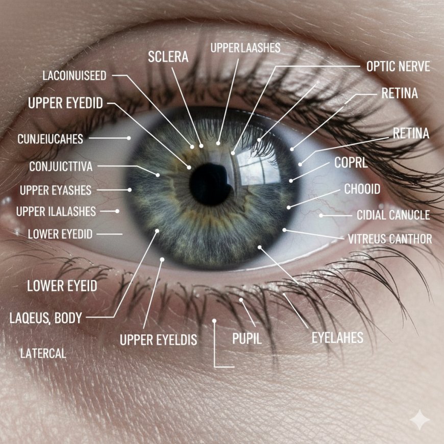
The eyebrows protect the eyes from sweat, dust, and sunlight. The eyelids protect the eye from injury and dryness and help spread tears over the surface. The eyelashes trap dust and foreign particles. The lacrimal apparatus includes the lacrimal gland and ducts which produce and drain tears, keeping the eye lubricated and protected. The conjunctiva is a thin transparent membrane lining the inside of the eyelids and covering the sclera. It also provides protection and lubrication.
Internal Anatomy of the Eye
The eyeball has three main layers. The outer fibrous layer includes the sclera and the cornea. The sclera is the white part of the eye. It is tough and maintains the shape of the eye while providing attachment for eye muscles. The cornea is the clear front part of the eye. It has no blood vessels and is responsible for bending light as it enters the eye. It is highly sensitive because of its rich nerve supply.
The middle vascular layer is called the uvea and includes the choroid, ciliary body, and iris. The choroid is rich in blood vessels and nourishes the retina while absorbing excess light to prevent reflections. The ciliary body contains ciliary muscles that control the shape of the lens and ciliary processes that produce aqueous humor. The iris is the colored part of the eye. It has a central opening called the pupil which regulates the amount of light entering the eye. It contains circular muscles that constrict the pupil and radial muscles that dilate it.
The inner nervous layer is the retina. It contains photoreceptor cells called rods and cones. Rods help with black and white vision and are highly sensitive in dim light. Cones help with color vision and sharp visual clarity and are concentrated in the fovea. The macula lutea is responsible for sharp central vision and the fovea is the point of highest visual acuity. The optic disc is also part of the retina. It is a blind spot because it contains no photoreceptors. It is the point where the optic nerve leaves the eye.
Chambers of the Eye
The eye has three chambers. The anterior chamber is between the cornea and iris and contains aqueous humor. The posterior chamber is between the iris and lens and also contains aqueous humor. The vitreous chamber is the largest chamber and is located behind the lens. It contains the vitreous body, a jelly-like substance that helps maintain the shape of the eyeball.
Lens
The lens is a transparent, biconvex structure responsible for accommodation. It can change its shape to focus light on the retina. The lens becomes thicker for near objects and thinner for distant objects. It is avascular and elastic.
Extraocular Muscles
The eye has six muscles that control movement. These include the superior rectus, inferior rectus, medial rectus, lateral rectus, superior oblique, and inferior oblique. Most of these muscles are controlled by the oculomotor nerve. The superior oblique is controlled by the trochlear nerve and the lateral rectus is controlled by the abducens nerve.
Physiology of Vision
Light enters the eye through the cornea, aqueous humor, pupil, lens, and vitreous body before reaching the retina. Most of the bending of light occurs at the cornea while the lens performs fine focusing. The lens changes its shape through accommodation. For near vision, the lens becomes thicker. For distant vision, it becomes flatter.
In the retina, rods and cones convert light into electrical signals. These signals pass to bipolar cells and then to ganglion cells. The axons of the ganglion cells form the optic nerve. These nerve signals travel through the optic nerve, optic chiasma, optic tract, lateral geniculate body, optic radiation, and finally to the visual cortex in the occipital lobe of the brain. The brain interprets these signals and produces the sense of vision.
Pupillary Reflex
The pupil changes size depending on the light. In bright light, the pupil constricts. In dim light, it dilates. This process is controlled by the autonomic nervous system.
Tear Formation
The lacrimal glands produce tears which clean, lubricate, and protect the eye. Tears drain through the lacrimal puncta, canaliculi, lacrimal sac, and nasolacrimal duct.
Common Eye Conditions
Some common conditions include myopia, hyperopia, astigmatism, cataract, glaucoma, conjunctivitis, retinopathy, and color blindness.
Short Questions (10)
1. What is the function of the cornea?
2. Define retina and name its two types of photoreceptor cells.
3. What is the role of the iris in vision?
4. Why is the optic disc called the blind spot?
5. What is aqueous humor and where is it found?
6. Explain the function of the lens.
7. What is the difference between rods and cones?
8. What is the function of the lacrimal gland?
9. Name any two extraocular muscles of the eye.
10. What is accommodation?
-Long Questions (5)
1. Describe in detail the anatomy of the human eye including all three layers of the eyeball.
2. Explain the physiology of vision from the entry of light into the eye to the formation of the final image in the brain.
3. Discuss the structure and functions of the retina. Add notes on rods, cones, macula, and optic disc.
4. Explain the role of the cornea, lens, aqueous humor, and vitreous humor in the refraction of light.
5. Describe the lacrimal apparatus and the mechanism of tear formation and drainage.
What's Your Reaction?
 Like
0
Like
0
 Dislike
0
Dislike
0
 Love
0
Love
0
 Funny
0
Funny
0
 Angry
0
Angry
0
 Sad
0
Sad
0
 Wow
0
Wow
0