


Wound Dressing and Management scenario based
This comprehensive guide cover appropriate dressing types and techniques for various wound care scenarios clear clinical actions and tips .
Complete Guide for Common Clinical
Scenarios
Wound care is a critical aspect of patient recovery. Selecting the right type of dressing and technique can significantly influence healing time, comfort, and infection prevention. Below, we explore common clinical wound care scenarios and provide evidence-based recommendations for dressing types and nursing interventions.
1. Venous Leg Ulcer – Daily Dressing and Compression Bandaging
Dressing: Hydrocolloid or foam dressing
Bandaging Technique: Multi-layer compression bandaging (e.g., four-layer compression system)
Rationale:
Venous leg ulcers result from chronic venous insufficiency, leading to poor circulation and edema. A foam or hydrocolloid dressing maintains a moist wound environment and absorbs moderate exudate. Compression therapy is essential to reduce venous hypertension and edema, promoting healing.
Ensure ankle-brachial pressure index (ABPI) is checked before applying compression to rule out arterial disease.
2. Diabetic Foot Ulcer – Moist Wound Healing
Dressing: Hydrogel or alginate dressing
Application: After cleansing the wound, apply the hydrogel directly onto the ulcer and cover with a secondary dressing. Secure gently with a non-adhesive wrap.
Rationale:
Hydrogels maintain a moist wound bed, promoting autolytic debridement and epithelialization—essential in diabetic wounds. Alginate can be used if exudate is heavy. Avoid adhesives directly on fragile diabetic skin.Offloading pressure using specialized footwear or foam padding is also critical.
3. Signs of Wound Infection During Dressing Change
Signs Noted: Purulent discharge, erythema, odor, warmth, increased pain
Immediate Actions:
Stop the dressing change
Notify the physician
Obtain a wound swab for culture and sensitivity
Document findings (odor, color, amount of drainage)
Apply a temporary clean dressing
Monitor for systemic infection (fever, chills)
Rationale:
Prompt identification and intervention can prevent worsening infection or sepsis. Documentation supports treatment planning and continuity of care.
4. Stage III Pressure Injury in Bedridden Patient
Dressing: Foam dressing with antimicrobial silver (if signs of colonization) or hydrofiber dressing
Considerations:
Depth and presence of tunneling
Amount of exudate
Risk of infection
Offloading pressure using a pressure-relieving mattress
Rationale:
Stage III wounds involve full-thickness tissue loss. Foam dressings provide cushioning and moisture balance. Silver-impregnated dressings help control bioburden.
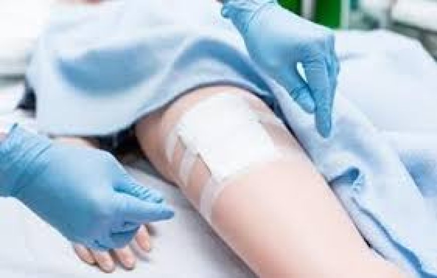
5. Post-Operative Abdominal Dressing Soaked with Serosanguinous Fluid
Management:
Remove the soaked dressing using aseptic technique
Inspect the wound for dehiscence or bleeding
Reinforce with sterile gauze and abdominal pads as needed
Document: color, amount, odor of drainage, patient condition
Notify surgeon if saturation recurs or increases
Rationale:
Serosanguinous fluid is normal post-op, but excessive saturation may indicate a complication. Timely action prevents skin maceration and infection.
6. Compression Bandage Causing Tingling and Coldness in Toes
Response:
Immediately remove or loosen the compression bandage
Assess circulation: capillary refill, pulses, skin color, and temperature
Re-evaluate bandage technique and pressure
Document findings and notify the healthcare provider
Rationale:
Tingling and coldness may indicate impaired circulation or nerve compression. Compression must be therapeutic but not constrictive.
Partial-Thickness Burns – Minimizing Pain During Dressing
Dressing: Non-adherent silicone dressing (e.g., Mepitel) or hydrogel sheets
Technique:
Cleanse gently with sterile saline
Apply a silicone dressing that does not stick to the wound
Use gauze as secondary dressing and secure with light wrap
Pre-medicate for pain if needed
Rationale:
Silicone dressings reduce trauma and pain during dressing changes while maintaining a moist healing environment.
8. Heavily Exudating Wound – Absorbent Dressing
Dressing: Calcium alginate or hydrofiber dressing (e.g., Aquacel)
Rationale:
These dressings absorb large volumes of exudate, help manage maceration risk, and promote autolytic debridement. They conform well to wound contours and require fewer changes, enhancing patient comfort.Always monitor surrounding skin for signs of moisture-associated damage.
9. Knee Laceration in a Child – Flexibility and Movement
Dressing: Non-adherent dressing with flexible adhesive (e.g., Tegaderm + non-stick pad)
Bandaging: Elastic cohesive bandage or tubular bandage
Rationale:
Children are active, so the dressing must move with the joint. Using a non-stick dressing under a transparent film allows visibility while promoting healing. The bandage must not restrict motion but should hold the dressing in place.
10. Caregiver Education for Pressure Ulcer Dressing at Home
Key Instructions:
Hand hygiene before and after dressing change
Use sterile or clean gloves depending on wound protocol
Clean the wound with normal saline or prescribed solution
Apply the correct dressing and avoid touching the wound bed
Observe for signs of infection: odor, pus, redness
Dispose of old dressings safely in sealed bags
Keep a wound care log for healthcare visits
Ensure offloading and frequent repositioning
Rationale:
Educating caregivers ensures continuity of aseptic technique and supports proper wound healing at home. Clear, written instructions help reduce errors and complications.
What's Your Reaction?
 Like
0
Like
0
 Dislike
0
Dislike
0
 Love
0
Love
0
 Funny
0
Funny
0
 Angry
0
Angry
0
 Sad
0
Sad
0
 Wow
0
Wow
0